Q1. ATPase enzyme needed for muscle contraction is located in
Solution
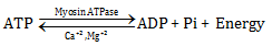
During muscle contraction, hydrolysis of ATP into ADP and inorganic phosphate occurs. The energy released during the process raises the meromyosin head to a high-energy state. The enzyme myosin ATPase catalyses the reaction in the presence of Ca2+ and Mg2+.
Q2. Draw a well-labelled diagram of the rib and rib cage.
Solution
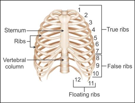
Rib and rib cage:
Q3. The membranous areas between the cranial bones of the foetal skull are called
Solution
Fontanelles comprise any of the soft membranous gaps between the incompletely formed cranial bones of a foetus or an infant. They allow for rapid stretching and deformation of the cranium as the brain expands faster than the surrounding bone can grow.
Q4. Name the embryonic layer from which the muscles originate.
Solution
Mesoderm
Q5. The
number of vertebrae present in the cervical, thoracic, lumbar, sacral and
coccyx regions, respectively, are
Solution
The vertebrae
are grouped into five categories:
Cervical
vertebrae: 7, present in the neck
Thoracic
vertebrae: 12, present in the upper back
Lumbar
vertebrae: 5, located in the abdomen
Sacral
vertebrae: 5, present in the pelvis
Coccygeal
vertebrae: 4, present in the vestigial tail
Q6. Distinguish between pectoral and pelvic girdles.
Solution
Pectoral Girdle
Pelvic Girdle
It consists of the scapula and the clavicle.
It consists of the coxal bones made by the fusion of the ilium, ischium and pubis.
It bears the glenoid cavity.
It bears the acetabulum cavity.
The two halves of the pectoral girdle are not joined to each other, but they are joined to the sternum on its either side.
The two halves of the pelvic girdles are joined to each other by the pubic symphysis.
Q7. Draw the cross-sectional view of a muscle showing muscle fibres and
bundles.
Solution
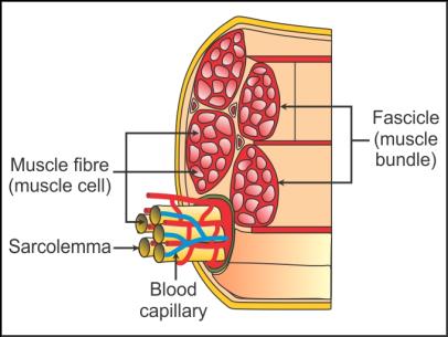
Cross-sectional view of a muscle showing muscle fibres and bundles:
Q8. Name the vertebra which articulates with the occipital condyles.
Solution
Atlas
Q9. Represent diagrammatically:
Structure of actin filament
Structure of myosin filament
Solution
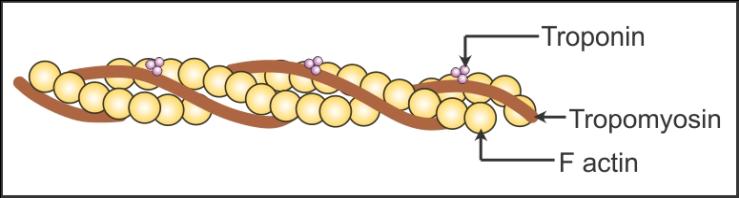
a. Structure of actin filament:
 b. Structure of myosin filament:
b. Structure of myosin filament:
Q10. How many bones are present in the front part of the skull which
protects the facial region?
Solution
14
Q11. Write any two special properties of muscles.
Solution
Extensibility and elasticity
Q12. Describe the synovial joint.
Solution
The synovial joint is characterised by the synovial cavity filled with the synovial fluid.
The synovial cavity is present between the articulating bones of the joint.
This arrangement and the presence of the lubricating fluid allow considerable movement.
Some examples are the knee joint and the ball and socket joint present between the glenoid cavity and head of the humerus.
Q13. Give any two examples of the synovial joints.
Solution
Two examples of the synovial joints are
Joint between the humerus and the pectoral
girdle
Knee joint
Q14. Which ions are stored by the sarcoplasmic reticulum in muscles?
Solution
Calcium ions are stored by the sarcoplasmic reticulum in muscles.
Q15. State the important functions of the vertebral column.
Solution
The functions of the vertebral column are as follows:
It protects the spinal cord.
It supports the head.
It serves as the point of attachment for the
ribs and the musculature of the back.
Q16. Define locomotion.
Solution
Voluntary movements which bring a change in place or location of the
organism are called locomotion.
Q17. In the pelvic girdle of man, A B, C, D and E, respectively, represent
Solution
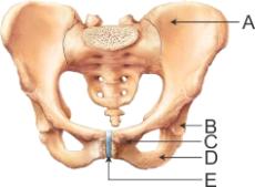
The pelvic girdle is located in the lower part of the trunk. The bones of the pelvic girdle shown in the diagram are
A - Ilium, B - Acetabulum, C - Pubis, D - Ischium, E - Pubic symphysis
Q18. Name the filaments found arranged parallel in a muscle fibre.
Solution
Myofibrils
Q19. Which bone articulates with the glenoid cavity?
Solution
The head of humerus articulates with the glenoid cavity.
Q20. What is a sarcomere?
Solution
Sarcomere is the functional region of the myofibril between two
successive Z lines.
Q21. Distinguish between fibrous joints and cartilaginous joints.
Solution
Fibrous Joints
Cartilaginous Joints
These
joints are immovable joints and do not allow any movement.
These
joints provide limited movements.
The
flat bones are fused end-to-end by dense fibrous connective tissues.
The
bones are joined by the cartilage.
Q22. Which one of the following is the correct pairing of a body part and the kind of muscle tissue which moves it?
Solution
Iris - Involuntary smooth muscles
Heart wall - Cardiac muscles
Biceps of upper arm - Striated muscle fibres
Abdominal wall - Smooth muscles
Q23. Which bone of the axial skeleton is made of 8 bones?
Solution
Cranium
Q24. Name the bones which form the rib cage.
Solution
The thoracic vertebrae, ribs and sternum form the rib cage.
Q25. Explain the structure of the myofibril.
Solution
Structure of Myofibril:
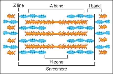 The myofibril is a unit filament of a muscle fibre.
It is made of two kinds of bands - dark band and light band.
A dark band is also known as an A or anisotropic band (myosin filament) and contains myosin, while the light band is also known as an I or isotropic band (actin filament) and contains actin.
Both the bands or filaments are arranged parallel to each other.
Actin filaments are thinner than myosin filaments and are hence also called thin and thick filaments, respectively.
At the centre of each I band is an elastic fibre called Z-line to which thin filaments are firmly attached.
The thick filaments in the A band are held together by an M line.
The portion of the myofibril between two successive Z lines is considered functional and is called a sarcomere.
In the resting state, the edges of thin filaments partially overlap the free ends of thick filaments on either side leaving the central parts of the thick filament non-overlapped; this is called the H zone.
The myofibril is a unit filament of a muscle fibre.
It is made of two kinds of bands - dark band and light band.
A dark band is also known as an A or anisotropic band (myosin filament) and contains myosin, while the light band is also known as an I or isotropic band (actin filament) and contains actin.
Both the bands or filaments are arranged parallel to each other.
Actin filaments are thinner than myosin filaments and are hence also called thin and thick filaments, respectively.
At the centre of each I band is an elastic fibre called Z-line to which thin filaments are firmly attached.
The thick filaments in the A band are held together by an M line.
The portion of the myofibril between two successive Z lines is considered functional and is called a sarcomere.
In the resting state, the edges of thin filaments partially overlap the free ends of thick filaments on either side leaving the central parts of the thick filament non-overlapped; this is called the H zone.
Q26. Name the substance which when accumulated in muscles causes fatigue.
Solution
Lactic acid
Q27. Represent diagrammatically the sliding filament theory.
Solution
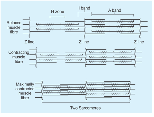
Sliding filament theory:
Q28. What are the bones of the palm called?
Solution
Metacarpals
Q29. Name the muscles present in the inner wall of the reproductive tract.
Solution
Smooth muscles
Q30. State the name for the plasma membrane of the muscle fibre.
Solution
Sarcolemma
Q31. Name the central hollow portion of the vertebra.
Solution
Neural canal
Q32. State the two major divisions of the human skeletal system.
Solution
Two major divisions of the human skeletal system are
Axial skeleton
Appendicular skeleton
Q33. Name the two bones of the pectoral girdle.
Solution
Scapula and clavicle
Q34. Describe the structure of actin filament.
Solution
The actin filament is made of two polymer F actins which are helically
wound to each other.
Each F actin is made of a monomer called globular or G actin.
Two filaments of a protein called tropomyosin run close to the F
actins throughout their length.
At regular intervals, a protein named troponin is distributed on the
tropomyosin.
In the resting phase, the active binding site for myosin present on
the actin filament gets masked by the troponin.
Q35. The largest muscle in the human body is
Solution
The largest muscle in the human body is gluteus maximus, which is also known as the buttock muscle. It is large because its main function is to keep the body upright.
Q36. Why are the ribs called bicephalic? What are true ribs?
Solution
Because the rib bones have two articulation surfaces on the dorsal side, they are called bicephalic.
The first seven pairs of ribs are dorsally attached to the thoracic vertebrae, and they are ventrally connected to the sternum. These ribs are called true ribs.
Q37. Name the cartilage which helps vertebrochondral ribs to join the
seventh rib.
Solution
Hyaline cartilage
Q38. Name the three bones which fuse to form the coxal bone.
Solution
Ilium, ischium and pubis fuse to form the coxal bone of the pelvic
girdle.
Q39. Name the three types of muscles based on their location.
Solution
The three types of muscles based on their locations are
Cardiac muscles
Skeletal muscles
Visceral muscles
Q40. Which pairs of ribs are called false ribs? Why?
Solution
The 8th, 9th and 10th pairs of ribs
are called false ribs.
These three pairs of ribs do not articulate directly with the sternum.
Instead they are joined to the 7th pair by the hyaline
cartilage.
Q41. Name the muscle involved in changes of body posture.
Solution
Skeletal muscles
Q42. Explain the following disorders:
Osteoporosis
Tetany
Muscular dystrophy
Solution
Osteoporosis: A decreased level of oestrogen is
the common cause. The bone mass is decreased due to which a risk of
fracture increases. It usually occurs in old individuals.
Tetany: Rapid spasms are experienced in muscles
due to a low calcium ion level in body fluids.
Muscular dystrophy: It is a genetic disorder. The
skeletal muscles degenerate progressively.
Q43. Represent diagrammatically the anatomy of a sarcomere.
Solution
Anatomy of a Sarcomere:

Q44. Name the muscular disorder which affects the neuromuscular junction or
motor-end plate.
Solution
Myasthenia gravis
Q45. Explain the contraction and relaxation of muscles by the sliding
filament theory.
Solution
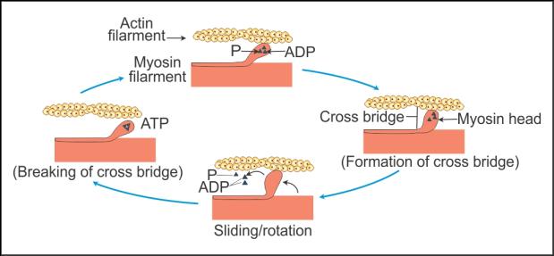
Muscle contraction is initiated by the signal sent by the central
neural system to the motor end-plate.
As soon as the motor end-plate receives the single, acetylcholine is
released at the end-plate which sets the action potential in the sarcolemma.
As the action potential spreads through the fibre, it causes the
sarcoplasm to release calcium ions.
Increase in the calcium level results in binding of calcium with the
troponin present on the actin filament.
The binding of calcium with the troponin unmasks the active binding site
for myosin which is present on the actin filament.
Myosin binds with the active site forming the cross-bridge. The energy
required for the binding of myosin is obtained from the hydrolysis of ATP.
Formation of the cross-bridge pulls the attached actin filaments
towards the centre of the A band, and the Z lines are pulled inwards,
resulting in the contraction of the sarcomere.
As soon as ADP and iP are formed from ATP, myosin goes back to its
relaxed state and the cross-bridge is broken.
At some point, calcium ions are pumped back to the sarcoplasmic
cisternae, which results in the masking of the active binding sites of myosin,
resulting in the relaxation of muscle fibre.
Q46. Represent diagrammatically the various stages of cross-bridge formation in muscles.
Solution
Stages of cross-bridge formation in muscles:
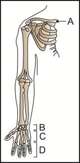
Q47. Study the given figure. Identify A, B, C and D. Write the number of
bones present in B, C and D.
Solution
A - Clavicle
B - Carpals (8 bones)
C - Metacarpals (5 bones)
D - Phalanges (14 bones)
Q48. Name the monomer of myosin filament.
Solution
Meromyosin
Q49. Name the cells which show amoeboid movement and are found in the human body.
Solution
Macrophages and leucocytes
Q50. Name the oxygen-storing pigments of muscles.
Solution
Myoglobin
Q51. Explain the structure of the pectoral girdle.
Solution
The pectoral girdle is made of the scapula and the clavicle.
The scapula is a large triangular bone which is located on the dorsal
part of the thorax between the second and the seventh ribs. It bears an
elevated ridge called the spine which extends as the expanded flat process
called the acromion.
The clavicle articulates with the acromion.
The scapula bears a depression called the glenoid cavity below the
acromion.
The head of the humerus of the forelimb articulates with the glenoid
cavity.
The clavicle is a long slender bone with two curvatures. It is also
called the collar bone.
Q52. Name the joints which do not allow any movement.
Solution
Fibrous joints
Q53. Name the hormone responsible for osteoporosis.
Solution
Oestrogen
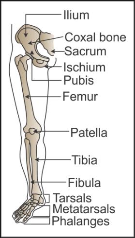
Q54. Represent diagrammatically the pelvic girdle and the bones of the hindlimbs.
Solution
Pelvic girdle and the bones of the hindlimbs:
Q55. Which cell organelle is abundantly present in aerobic muscles?
Solution
Mitochondrion
Q56. Name the cavity to which the thigh bone articulates.
Solution
Acetabulum
Q57. Distinguish between red and white muscle fibres.
Solution
Red Muscle Fibre
White Muscle Fibre
Myoglobin
content is high.
Myoglobin
content is low.
Contain
more number of mitochondria.
Contain
less number of mitochondria.
Q58. Name the junction between a motor neuron and a sarcolemma.
Solution
Neuromuscular junction or motor end-plate
Q59. State the different regions of the vertebral column and the number of
bones present in each region.
Solution
Regions of the vertebral column
Number of bones present
Cervical region
7
Thoracic region
12
Lumbar region
5
Sacral region
1, fused
Coccygeal region
1, fused
Q60. Differentiate between skeletal muscles and visceral muscles.
Solution
Skeletal Muscles
Visceral Muscles
They
show the presence of striations and are hence called striated muscles.
Striations
are absent, and they are hence called smooth muscles.
Activities
are under the control of the nervous system; hence, they are voluntary
muscles.
Their
activities are not under the control of the nervous system; hence, they
are called involuntary muscles.
They
are responsible for locomotory actions and body posture.
They
are responsible for the movement of food through the digestive tract
or the movement of gametes in the genital tract.
Q61. Which parts of myosin filament act as ATPase?
Solution
The globular head of the myosin monomer acts as ATPase.
Q62. What is the technical term used for the knee cap?
Solution
Patella
Q63. Name the tissue which holds all the muscle bundles together in a muscle.
Solution
Fascia
Q64. State the symptom of gout.
Solution
Symptom of gout:
Inflammation of joints
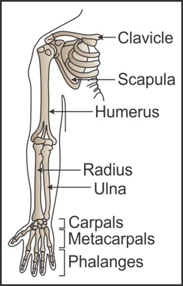
Q65. Represent diagrammatically the pectoral girdle and the bones of the forelimbs.
Solution
Pectoral girdle and the bones of the forelimbs:
Q66. Describe the structure of myosin as a contractile protein.
Solution
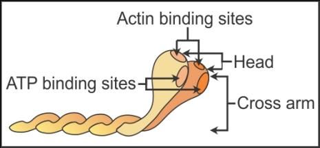
Structure of myosin filament:
Each myosin filament is made of monomers of protein called meromyosin.
Each meromyosin has two regions - heavy meromyosin (HMM), which is a globular head with a short arm, and light meromyosin (LMM), which is the tail.
The globular head acts as an active ATPase, and it also has the binding site for actin.
The globular head and the short arm project outwards at a regular distance and angle from each other and from the surface of the myosin filament. This structure is called the cross-arm.
Q67. Name the neurotransmitter released at the neuromuscular junction.
Solution
Acetylcholine
Q68. Name the two proteins present in the myofilaments of muscles.
Solution
Actin and myosin
Q69. How many bones are present in the human skeletal system?
Solution
206 bones
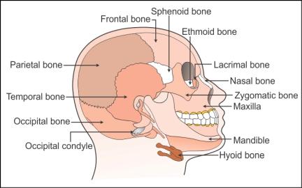
Q70. Draw a well-labelled diagram of the human skull and label any 10 parts.
Solution
Human skull:
Q71. Name the bone present at the base of the buccal cavity.
Solution
Hyoid bone is present at the base of the buccal cavity.
Comments
Post a Comment